
Diagnosing a traumatic brain injury is no longer limited to what shows up on a standard brain scan. Today, doctors can use blood tests, advanced imaging, and detailed symptom records to get a clearer picture after someone suffers a head injury. This matters because even a mild traumatic brain injury, like a concussion, can affect memory, concentration, balance, sleep, and overall brain function, even when a CT scan looks normal. These tools can also help provide medical evidence in a California injury claim. No test, however, replaces a clinician’s judgment or decides a legal case.
The Problem: “Invisible” Brain Injuries
CT scans are built to find urgent problems such as bleeding, skull fracture, swelling, or a midline shift. They are essential when those dangers are suspected, but they are not designed to show every subtle brain injury. The Centers for Disease Control and Prevention explains that a scan is not required to identify mild traumatic brain injuries or concussions. A negative CT scan therefore does not mean that every symptom is imaginary or unrelated to the injury.
That gap creates real difficulty for patients with suspected TBI after motor vehicle accidents, falls, or other head injuries. A person may appear well while experiencing headaches, slowed thinking, light sensitivity, cognitive difficulty, or mood changes. Insurers may focus on a normal scan or on the word “mild” and ignore the injury’s functional impact. Clear medical follow-up, consistent symptom reporting, and evidence of changes at work or home help show what the injury has actually done.
How Traumatic Brain Injury Diagnosis Is Changing
TBI diagnosis used to rely mainly on Glasgow Coma Scale scores, CT scans, and observation over the first 24 to 48 hours. If you looked “okay” on those measures, many providers assumed you were.
Over the last decade that picture has changed. Advances in neuro-technology have improved TBI diagnosis at every stage — from the first minutes after a head injury through months of follow-up. Researchers have developed blood tests that detect specific proteins released by damaged brain cells, and imaging techniques that reveal structural and functional changes invisible to older scans.
Large multi-center observational studies like the TRACK-TBI Study have shown that many TBI patients with normal CT scans still have brain injuries detectable on MRI or in blood-based biomarkers. A negative CT scan no longer means “no injury.”
These tools do not replace a physician’s judgment. They add objective data points that help doctors confirm or rule out brain injuries with greater confidence. For a person in California considering a claim, these tests can later help show that the original injury was real, not merely “subjective complaints.”
Blood Tests for Brain Injury
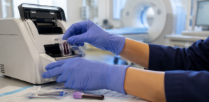
A blood test for brain injury looks for specific proteins released from damaged brain tissue into the bloodstream after head trauma. When brain cells are injured, they leak these protein biomarkers into the blood, where they can be measured.
Two biomarkers lead the way:
- GFAP (glial fibrillary acidic protein) is released from injured astroglial cells. It becomes detectable within about one hour of injury, peaks around 20 hours, and can remain elevated for days. GFAP levels can indicate the severity of brain injury and can detect injuries that CT scans miss.
- UCH-L1 (ubiquitin C-terminal hydrolase L1) comes from damaged neurons. It rises sharply in the first hours but drops toward baseline within about 24 hours.
A peer-reviewed review of these biomarkers confirms that GFAP and UCH-L1 have distinct time courses: UCH-L1 peaks early and fades fast, while GFAP has a wider detection window, making it especially useful when patients arrive at the hospital hours after injury.
Abbott’s i-STAT TBI whole blood test, cleared by the FDA in 2024, measures GFAP and UCH-L1 from a simple blood draw and returns results in about 15 minutes at the patient’s bedside — without the centrifuge step earlier plasma-based versions required. According to the FDA clearance summary, the test showed a high negative predictive value for ruling out CT-visible intracranial injury, which is its primary clinical use: helping emergency clinicians decide who does not need a CT scan.
For longer-term monitoring, ultrasensitive laboratory platforms can detect NfL (neurofilament light), a protein released from damaged axons that rises more slowly and can stay elevated for weeks to months. NfL is particularly useful for tracking ongoing injury and for predicting longer-term cognitive outcomes.
Biomarkers can also help distinguish the acute and chronic phases of TBI, which helps doctors — and attorneys — understand where a patient sits on the recovery timeline.
Advanced Imaging and AI
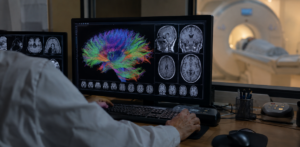 Standard CT scans detect emergencies such as major bleeding, fractures, and midline shift. Conventional MRI provides greater soft tissue detail but still misses many subtle brain injuries. Advanced imaging techniques go further.
Diffusion Tensor Imaging is an advanced MRI technique that tracks the movement of water molecules along white matter tracts — the neural pathways connecting different brain regions. When axons are stretched, torn, or damaged, water diffusion patterns change. DTI can detect these changes even when standard brain scans look completely normal, particularly in the corpus callosum and other critical white matter tracts.
A large review of approximately 325 studies found that about 70% of DTI studies reported associations between DTI measures and clinical outcomes, including cognition and symptoms.
Functional MRI maps changes in brain activity by measuring blood flow during cognitive tasks. It can show, for example, that a patient’s brain works harder than normal to perform a simple memory task, or that certain networks do not activate at all. fMRI is more expensive and less standardized than DTI, but it offers insight into brain function that structural scans cannot.
Quantitative EEG, or qEEG, measures electrical signals from the scalp and compares them with normal patterns to detect abnormal brain activity after injury. Advanced EEG methods can also measure how the brain processes information and sustains attention. Together, these approaches help doctors see both the brain’s structure and how it works.
Artificial intelligence is entering the field as well. Machine learning models can analyze CT and MRI scans to flag small bleeds, microstructural changes, or injury patterns that are easy to overlook under heavy clinical workload. Deep learning systems trained on thousands of prior scans can assist in identifying traumatic brain injury.
AI will not replace a radiologist. It functions as a second set of eyes, speeding up and supporting detection. Any imaging decision should be made with your treating physicians. This article is for informational purposes only and is not medical advice.
Why This Matters for Your Injury Claim
To prove a brain injury, you need evidence linking the event to your diagnosis and showing how your symptoms affect daily life. Getting checked quickly, obtaining biomarker results, having a specialist review your scans, and keeping a consistent treatment record all provide evidence alongside your own account. Work and school records, and observations from family members, can show changes that a short doctor’s visit might miss.
Technology can help counter an insurer’s “you look fine” argument, particularly where a CT scan does not show a mild brain injury. But a biomarker result or advanced scan alone does not establish who was at fault, what caused the injury, or what a claim is worth. What matters is the full record: when symptoms started, your health history, expert opinions, and proof of lost income or reduced independence.
If someone else may be responsible, our traumatic brain injury lawyer can review your medical and accident records, address insurer disputes about an “invisible” injury, and explain how the claim process works in California. Because presentation depends on the specific facts, an attorney should review how these technologies apply to your situation.
What to Do If You Have a Brain Injury
- Get evaluated promptly. Go to an emergency room or urgent care after any significant head impact, even if you did not lose consciousness. Many brain injuries occur without a dramatic event. Tell the doctor exactly what happened.
- Follow up if symptoms continue. If headaches, confusion, memory problems, or mood changes persist in the days and weeks after injury, see your primary care doctor or a neurologist. Ask about blood tests and whether advanced imaging is appropriate.
- Keep copies of everything. Save all medical records, imaging reports (CT, MRI, DTI), discharge instructions, and any blood test results. These documents are the foundation of both your care and any future claim.
- Do not downplay symptoms. When talking to insurers or doctors, or filling out forms, describe honestly how the injury affects work, school, driving, sleep, and relationships. Minimizing symptoms now can undermine your case later.
- Consult a traumatic brain injury lawyer. Understand your rights, learn how diagnostic tools may support your claim, and avoid mistakes in recorded statements or releases. An attorney can coordinate with your medical team to preserve evidence.
Frequently Asked Questions
Featured Tech Merges TBI FAQs
Is there a blood test that can diagnose a concussion or mild TBI?
FDA-cleared blood tests measuring GFAP and UCH-L1 help emergency doctors decide who is at higher risk for brain injury and who may need imaging. They are not a simple yes-or-no “concussion test.” They are used primarily in the first hours after injury and represent one piece of information alongside symptoms, exam findings, and imaging. If you did not receive a blood test at the time of injury, other evaluations may still help document a brain injury later.
Does a normal CT scan mean I don’t have a brain injury?
No. A normal CT scan rules out major problems like large bleeds, skull fractures, or dangerous swelling, but it cannot detect most microscopic or diffuse axonal injuries associated with mild TBI. Research shows that many people with negative CT scans still have abnormalities on MRI or elevated blood biomarkers — and still have real symptoms. If you have ongoing problems after a head injury, follow up with a doctor even if your original CT was “normal.”
Can advanced imaging, such as DTI or functional MRI, help my legal case?
Sometimes. DTI and functional MRI can provide objective evidence of changes in brain structure or function that support a TBI diagnosis. Whether that evidence is admitted depends on the findings, the experts involved, and how the court applies California’s rules for scientific evidence. Some California trial courts have admitted DTI evidence, but no California appellate decision has settled the question, and defense counsel commonly challenges it — both on whether the technique is generally accepted for diagnosing TBI and on how the images are processed and interpreted. Admissibility is decided case by case. Discuss cost, benefit, and timing with both your treating physician and your attorney.
Will my insurance pay for advanced brain imaging or biomarker tests?
Coverage varies by health plan, provider network, and the medical necessity determined by your treating physician. Some plans cover DTI or specialized blood tests when ordered by a neurologist for clinical reasons; others do not. Talk with your doctor and insurer in advance. Legal strategy should not drive medical decisions — tests should be ordered because they help guide your care.
How long do I have to file a brain injury lawsuit in California?
Generally, California gives injured people two years from the date of injury to file a personal injury lawsuit under CCP 335.1. Shorter deadlines apply in some situations, including claims against government entities, which may require an administrative claim within six months. Missing the deadline — even while still undergoing testing or waiting for imaging results — can permanently end your right to pursue compensation. Our page on the deadlines that apply to injury claims covers the common exceptions. Do not wait for every test result before speaking with a lawyer.
Suffering After a Brain Injury? Talk to the Law Offices of Pius Joseph
Living with an “invisible” brain injury is exhausting, especially when friends, employers, or insurers do not understand what you are going through. The Law Offices of Pius Joseph works with medical professionals, imaging specialists, and emerging blood-test evidence to help document traumatic brain injuries in California claims.
If you or someone you love is dealing with symptoms after head trauma, do not wait for the filing deadline to pass. Schedule a free consultation to learn how these developments in medical technology may support your case.